Master of Biotechnology Student Builds Specialized Skills in Molecular and Cellular Imaging Facility
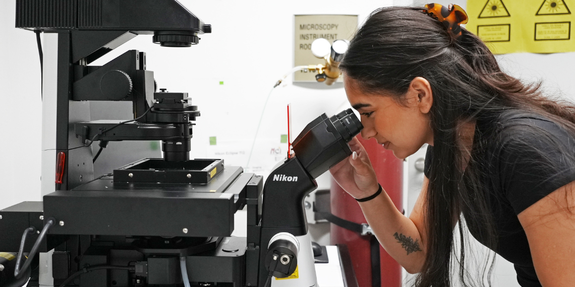
Soft cheese and peppers. Nanoparticles and nanowires. Bacteria and bacteriophages. Every day, a diverse assortment of samples from across the University of Guelph arrives at the Molecular and Cellular Imaging Facility, where Master of Biotechnology student Mojda Rashidi prepares them for a closer look.
Working in the facility this summer, Mojda prepares, processes and catalogues samples, while gaining hands-on experience with sophisticated research equipment, from electron microscopes and cell-sorting instruments to sample preparation tools such as sputter coaters and microtomes.
She also helps researchers determine the best imaging techniques for their project and provides training to students, postdoctoral researchers, and faculty alike on both light and scanning electron microscopes.
 On any given day, she could be meeting with researchers working in fields as diverse as physics, engineering, agriculture and microbiology.
“When I’m training someone, I’ll ask them about their research and what the imaging is for, just out of curiosity,” she says. “It's always really interesting to hear the different paths people are taking.”
The two most powerful microscopes that Mojda uses in the imaging facility, the scanning and transmission electron microscopes, provide incredibly detailed, high-resolution images needed for research across the University.
The scanning electron microscope reveals the three-dimensional structure of a sample’s surface, while the transmission electron microscope passes electrons through ultra-thin sections of a sample, revealing internal structures such as cell membranes and organelles. The scanning electron microscope can magnify up to about 100,000 times, while the transmission electron microscope magnifies up to 300,000 times.
“I remember the first time I sat down and looked at how clear the images were, how clearly you can see the cell walls of bacteria, for instance. It makes you feel kind of small, even though what you’re looking at is small,” says Mojda. “It’s crazy what you can see through a microscope.”
Preparing samples for the scanning electron microscope is a long, multistep process. Because these microscopes operate under a vacuum, hydrated samples must first be chemically fixed, gradually dehydrated, dried using a critical point dryer, and finally coated with a thin layer of gold before they can be imaged. The process can take up to two days.
Preparing samples for the transmission electron microscope takes less time but requires a steady hand. Thin sections or tiny droplets of material are placed on delicate copper grids, then washed and stained before imaging.

As part of her placement, Mojda is also contributing to research in Dr. Cezar Khursigara's lab on Pseudomonas aeruginosa, an antibiotic-resistant bacterium that frequently infects the lungs of people with cystic fibrosis. The research team is testing whether disabling specific genes in the bacterial biofilm would make the bacterium more vulnerable to treatments like antibiotics. Mojda uses scanning, transmission electron and light microscopy to examine how the genetic changes alter the bacteria’s structure.
While Mojda originally imagined a career in the pharmaceutical industry, the placement has introduced her to the wide range of careers in research.
"My main goal was to build transferable skills that I could take into my future career," she says. "Now I can see so many different paths."
