Displaying Results: [21 - 30] of 45
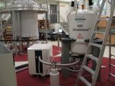
Dynamic Nuclear Polarization (DNP) NMR provides unparalleled sensitivity for membrane protein and other solid-state NMR samples. Upon addition of a stable radical species into the sample, microwaves are used to transfer polarization from the radical’s unpaired electron(s) to the nuclei of the sample of interest, providing a 30- to 40-fold boost in signal intensity, resulting in a time savings of 900- to 1600-fold over conventional NMR spectroscopy. This system is also equipped with a low-temperature MAS unit allowing for conventional MAS-NMR down to 100 K.
Facility: NMR Centre
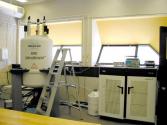
Offering the highest resolution and excellent sensitivity, this spectrometer is the crown jewel in the NMR Centre's inventory. This instrument is primarily used to study the structure and function of membrane proteins, with secondary focusses including the high-sensitivity detection of quadrupolar nuclei and high-resolution applications to small molecules and soluble proteins. The spectrometer is equipped with 4 RF channels and 3 high power RF amplifiers.
Facility: NMR Centre
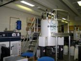
With a high-sensitivity cryoprobe, this instrument offers the highest signal-to-noise of all systems in the NMR Centre. This instrument is primarily used in metabolomics studies, as well as to study both isotopically enriched and natural abundance samples of proteins, nucleic acids, and carbohydrates, providing answers to questions of biological structure and function. The spectrometer is equipped with 4 RF channels and 4 RF amplifiers, allow simultaneous pulsing of 1H, 13C, 15N, and 2H nuclei.
Facility: NMR Centre
This "jack-of-all-trades" instrument is used to study solid and solution-state samples of chemicals, materials, and biological samples. A unique feature of this instrument is its two high-band and two low-band RF channels, allowing collection of 19F spectra with 1H decoupling (and vice-versa) as well as 13C spectra with simultaneous 19F/1H decoupling. Other broadband probes (both solids and liquids) allow for high-resolution detection of virtually any nucleus, while a High Resolution Magic Angle Spinning probe (HRMAS) is available to study solids and semi-solids with a high degree of internal motion.
Facility: NMR Centre
This versatile solid-state spectrometer is primarily used to study the properties of materials, polymers, and chemicals. This spectrometer is equipped with 4 RF channels and 3 high power RF amplifiers, and is equipped with gradient accessories, including a triple-axis gradient probe collar for gradient MAS studies and a Z-axis high power (60 A) gradient probe for diffusion studies.
Facility: NMR Centre
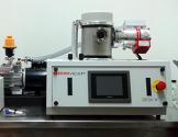
The Denton Desk V TSC is a high resolution unit suitable for oxidizing and non-oxidizing metals.
Type: Ancillary
Facility: Molecular and Cellular Imaging
The Denton DCP-1 is a Critical Point Dryer for preparing SEM samples.
Type: Ancillary
Facility: Molecular and Cellular Imaging
A versatile, high-performance instrument with three modes (high vacuum, low vacuum and ESEM) to image a wide range of samples, with or without preparation. Equipped with a Quorum PP3010 Cryo-Unit to freeze, fracture, etch and sputter coat temperature sensitive samples.
Type: Electron Microscopes
Facility: Molecular and Cellular Imaging
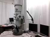
A 200kV field emission TEM that is used for cryo-temperature, low dose operation and atomic resolution.
Type: Electron Microscopes
Facility: Molecular and Cellular Imaging
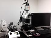
An upright Leica DM 5000B connected to a Hamamatsu Orca-Flash4.0 digital camera and Volocity™ imaging software.
Type: Optical Microscopes
Facility: Molecular and Cellular Imaging


