

Columnaris disease in rainbow trout
Hein Snyman, Lowia Al-Hussinee, Lisa Ledger, Hugh Cai
Animal Health Laboratory, University of Guelph, Guelph, ON.
AHL Newsletter 2024;28(3):16.
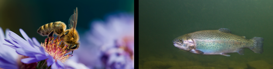
Flavobacterium columnare is the causative agent of “columnaris disease”, a significant ulcerative pathogenic bacterial infection that affects freshwater fish species. It is of particular economic significance in freshwater aquaculture, where the disease can result in devastating losses, especially in rainbow trout (Oncorhynchus mykiss), tilapia (Oreochromis sp.), and catfish (Ictalurus sp.).
The disease was first described in the early 1900s, and the causative bacterium has since then undergone numerous name changes, initially starting out as Bacillus columnaris and having been named due to its tendency to form column-like masses/colonies of bacteria when necrotic surface debris was reviewed on cytological examination (Fig. 1). Following that, the bacterium quickly transitioned through a variety of names, including Chondrococcus columnaris, Cytophaga columnaris, and Flexibacter columnaris, before finally settling on Flavobacterium columnare.
Although “columnaris disease” was historically thought to be caused by this singular species, more recent genetic work has resulted in the identification of four discrete genetic groups referred to now as “columnaris-causing bacteria (CCB)”, with these groups also exhibiting some specific species associations:
F. columnare (most common isolate in rainbow trout)
Flavobacterium covae sp. nov. (most common isolate in catfish)
Flavobacterium oreochromis sp. nov (most common isolate in tilapia)
Flavobacterium davisii sp. nov. (multiple different species)
Early infections can be non-specific with fish initially presenting with decreased feed intake, lethargy, a tendency to congregate or swim closer along the water surface, and increased opercular movement (gilling activity). Clinical symptoms quickly transition to the more characteristic signs of this disease which include patchy skin and fin discolouration with worsening erosion and eventual deep ulceration (Fig. 2). Although any external surface can be affected, changes are often most prominent along the dorsal fin and trunk, but other commonly-affected areas include the mouth/lips and gill arches. Histologically, lesions tend to be quite necrotizing with ulcerated surfaces and necrotic tissue being widely covered and permeated by dense mats of pale basophilic filamentous bacteria (Fig. 3, 4).
Mortality is usually associated with derangement of osmotic homeostasis following the loss of the integrity of the skin, however, hypoxia associated with significant loss of functional gill tissue can also contribute. Surface abrasion or pre-existent cutaneous injuries can play a significant role in the initial colonization by the bacterium, while stressors such as water temperature, low dissolved oxygen, or other aberrant aqueous environmental factors tend to initiate outbreaks and worsen disease progression. Flavobacteria are common aqueous environmental organisms, and as such, environmental contamination can significantly drive continued horizontal spread between fish.
Diagnosis of columnaris disease is based on a combination of clinical signs, wet mount cytology, gross and histological lesions, and positive bacterial isolation and identification (Fig. 5). However, specialized culture media is required for the isolation of Flavobacteria, and its availability is usually limited to laboratories that routinely deal with aquatic animal submissions. Given that isolation can sometimes also be difficult, the AHL has also recently validated a PCR assay for the identification of F. columnare in tissue samples (e.g., gill, skin, internal organs) which can further aid in confirmation. As with all PCR tests, positive results must be interpreted in conjunction with clinical signs, biopsy wet mount examination, and compatible gross and histological changes. Since initiating the F. columnare PCR assay in April 2024, we have tested 17 cases by PCR. Preliminary data shows that most cases have consistent results among the different tests, and that F. columnare PCR-positive results were significant even when the Ct value was as high as 40 (Table 1). AHL
Table 1. Comparison of F. columnare PCR with histology, gill and skin biopsy and isolation (NBP = No bacterial pathogens; NBG = No bacterial growth; ND = No data).


Figure 1. Wet mount cytology from the skin of a rainbow trout affected with columnaris disease exhibiting column-like stacked mats of slender filamentous bacteria (arrows).

Figure 2. Rainbow trout fish with cutaneous ulceration along the lateral body wall, characteristic of columnaris disease (arrow).

Figure 3. Histopathology of gill with regional necrosis of the gill filaments and abundant necrotic debris (arrows). H&E stain.

Figure 4. Higher magnification of the gill with necrotic debris being widely permeated by a dense meshwork of filamentous bacteria (arrows). H&E stain.

Figure 5. Bacterial culture plate morphology of a F. columnare colony exhibiting characteristic flat rhizoid structure with irregular margins. Also note the characteristic yellow pigmentation of the colony that can also afford yellow tissue discolouration in affected fish.
References
1. Smith PA, Elliot DG, Bruno DW, Smith SA. Skin and Fin Diseases. In: Fish Diseases and Medicine, 1st ed. Smith SA, ed. CRC press. 2019;5:108-110.
2. Bruno DW, Noguera PA, Poppe TT. Bacterial Diseases. In: A Colour Atlas of Salmonid Diseases, 2nd ed. Springer, 2013;6:84.
3. Benjamin R, et al. The fish pathogen Flavobacterium columnare represents four distinct species: Flavobacterium columnare, Flavobacterium covae sp. nov., Flavobacterium davisii sp. nov. and Flavobacterium oreochromis sp. nov., and emended description of Flavobacterium columnare. Syst Appl Microbiol. 2022;45(2):126293.