RUMINANTS
Bovine astrovirus – emergence of a novel viral encephalitis in cattle
Maria Spinato, Andrew Vince, Hugh Cai, Davor Ojkic
In May (steer A) and June (steer B) 2016, the AHL received fresh and formalin-fixed samples of brain from 2 steers located in separate feedlots, each comprised of 25-50 animals. Both steers had been found dead after exhibiting neurologic signs for several days. In steer B, tonic-clonic seizures were elicited by stimulation, and repetitive licking and chewing movements were also described. Prior to laboratory testing at the AHL, brain samples from both animals were submitted for Rabies virus fluorescent antibody testing by the Canadian Food Inspection Agency; both tested negative.
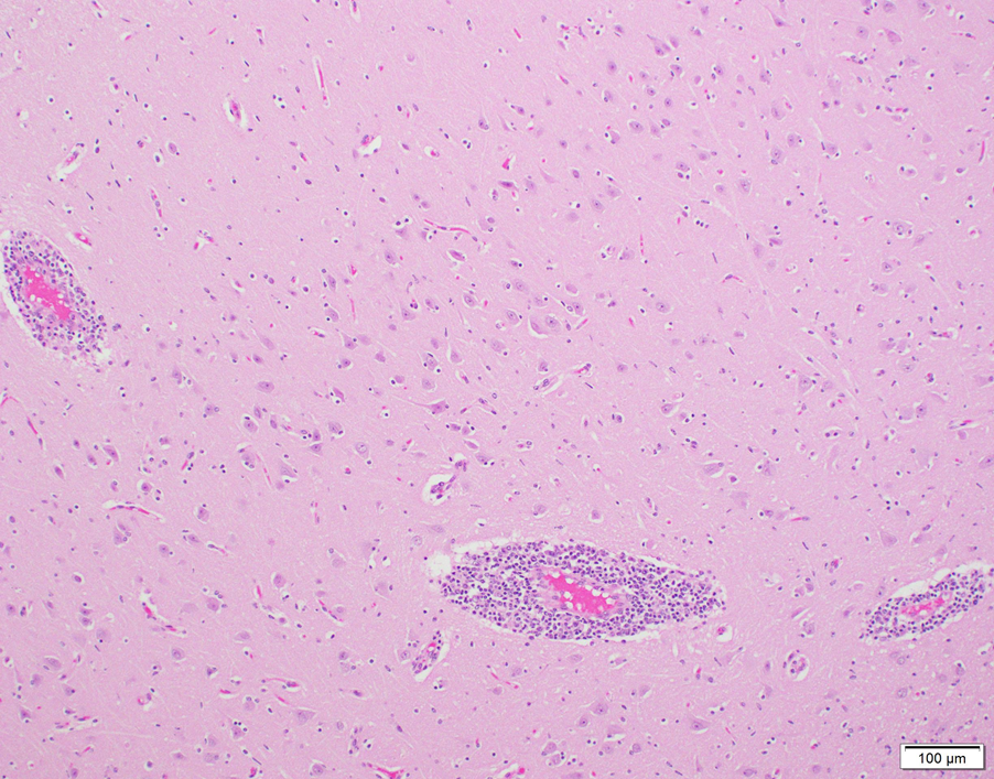
Histologic evaluation of brain revealed generalized, broad perivascular cellular cuffs extending from the cerebral cortex into the brainstem. Leukocytic cuffs expanding Virchow-Robin spaces were comprised of 3-8 layers of lymphocytes with occasional admixed histiocytes, rare neutrophils, and plasma cells (Fig. 1). Mild focal microgliosis and rare axonal spheroids were also observed in some sections. Regional nonsuppurative meningitis was present; vasculitis was not observed.
Based on the histologic lesions and the negative rabies test result, listerial meningoencephalitis and viral infections such as Bovine herpesvirus 1 and Ovine herpesvirus 2 (OvHV-2, Sheep-associated malignant catarrhal fever of cattle virus), were considered to be possible etiologies. Immunohistochemical staining for Listeria monocytogenes in paraffin-embedded sections of brain was negative in both cases, as were PCR tests for BoHV-1 and OvHV-2 in fresh sections of brain (OvHV-2 PCR performed by Prairie Diagnostic Services, Saskatoon, SK).
Idiopathic nonsuppurative meningoencephalitis is an occasional default final diagnosis in cattle with neurologic disease, given that a cause cannot be identified despite testing for a wide range of viruses, bacteria, and protozoa. Because of recent reports from Europe and the US of a novel astrovirus detected in cases of bovine nonsuppurative encephalitis, samples of brain from these 2 steers were tested for astrovirus by PCR. A RT-PCR specific for mammalian astrovirus1 was positive in both cases. The 385 bp PCR products were sequenced and demonstrated 95% homology with the US bovine astrovirus NeuroS12, and 93% homology with the Swiss bovine astrovirus CH13/NeuroS1 isolates3.
To our knowledge, this is the first confirmed report of bovine astrovirus-associated encephalitis in Canadian cattle.
It is suspected that neurotropic astroviruses have been circulating in global cattle populations for several decades. Swiss researchers who performed in situ hybridization on paraffin-embedded brain from cattle diagnosed with idiopathic encephalitis found astrovirus RNA in a high proportion of samples. However, viral association with histologic lesions was inconsistent, and the authors could not conclusively rule out a bystander role for this virus.4 Infection studies would assist in confirming the neurovirulent potential of bovine astrovirus, but have not been performed to date because the virus has yet to be isolated in cell culture.
Further work is in progress at the AHL to examine the association of bovine astrovirus and cases of idiopathic nonsuppurative encephalitis in Ontario cattle. AHL
References
- Mittelholzer C, et al. Molecular characterization of a novel astrovirus associated with disease in mink. J Gen Virol 2003;4:3087-3094.
- Li L, et al. Divergent astrovirus associated with neurologic disease in cattle. Emerg Infect Dis 2013;19:1385-1392.
- Bouzalas IG, et al. Full-genome based molecular characterization of encephalitis-associated bovine astrovirus. Infect Genet Evol 2016;44:162-168.
- Selimovic-Hamza ,et al. Detection of astrovirus in historical cases of European sporadic bovine encephalitis, Switzerland
 -ansi-language:en-CA;mso-ligatures:none'>
Figure 1. Non-suppurative perivascular cuffing in bovine brain in a case of astrovirus-associated encephalitis
Sign up for the Adult Small Ruminant Mortality Project!
Maria Spinato, Jocelyn Jansen, Paula Menzies, Andria Jones-Bitton

The AHL, OVC, and OMAFRA have received funding to conduct a joint study investigating adult small ruminant mortalities. Sick or dead adult sheep and goats are rarely sent to a laboratory for complete postmortem examination, and veterinarians infrequently perform postmortems on farm. However, there is value in knowing why an adult small ruminant died, especially during the current rapid expansion of small ruminant operations in Ontario.
The objectives of this study are:
- To determine why adult sheep and goats are dying on-farm.
- To determine if a web-based postmortem information and case submission system can be used to increase the usefulness of on-farm postmortems.
- To determine if better disease diagnoses can increase discussions between producers and their vets, so that they can create sound flock/herd health and biosecurity plans.
The project will fund:
- tissue collection kit and shipping fee,
- AHL laboratory test fees,
- compensation for veterinarian performing the on-farm postmortem.
In return, the veterinarian will submit:
- a complete set of fresh and formalin-fixed tissues, as per project protocol,
- a set of 6 postmortem photographs that are uploaded to the website,
- postmortem submission form with all required information, also uploaded to the website.
Funding support is being provided by the OMAFRA-University of Guelph Partnership KTT Program and the Ontario Animal Health Network. Thanks to the generosity of these agencies, ~170 postmortems of adult sheep and goats will be performed on farm over the next 12-16 months. A standardized diagnostic test panel has been developed to check for the diseases of greatest importance to the small ruminant industry.
Interested in participating?
For more information, contact the project administrators at sr.mort@uoguelph.ca or log onto the project web-site at https://www.uoguelph.ca/srmort/index.php/veterinarian/ AHL


