Protein Puzzles

U of G researchers follow the unfolding of cellular proteins to find out what makes them stable.
For more than 50 years, the protein-folding problem has remained unsolved. In living things, proteins begin as long strings of amino acids that fold themselves into specific 3D shapes. The 3D shape of a folded protein dictates the function of that protein. Many aspects of protein folding are not well understood, such as how the original amino acid chain causes a specific folding pattern. This is especially true for proteins which are embedded into cell membranes, the surface of cells. Such membrane proteins are partially water-soluble and partially fat-soluble, making them difficult to study. Common methods for studying protein structure require removing the membrane protein from its natural state, the cell membrane, which often alters its 3D structure. Less than 0.5% of known membrane proteins’ 3D structures have been determined, and we know little about how these structures are made by cells and what holds them together. Expanding upon this knowledge could have implications for future scientific applications that could be related to any living thing, including humans.
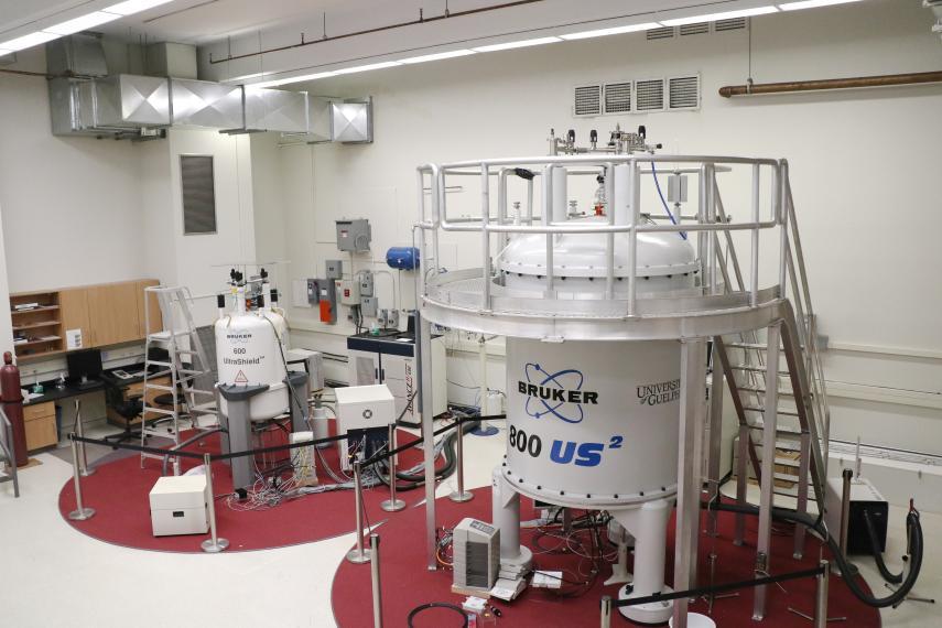
A research team led by University of Guelph physics professors Leonid Brown and Vladimir Ladizhansky found that membrane protein 3D structure stabilization factors could be determined by using solid-state NMR (ssNMR) to observe how the protein unfolds. The research team isolated a membrane protein from bacterial cells and placed it into a lipid (fat) mixture that mimics a cell membrane. They applied increasing amounts of heat to unfold the test protein step by step. To analyze each step they combined ssNMR with a technique called hydrogen/deuterium exchange to visualize the specific parts of the protein that have become exposed to water after a certain amount of heat was added (as it unfolds, more of the protein is exposed). By following the changes in protein structure as it unfolds, researchers were able to determine the main factors of its stability.
“Combining ssNMR and hydrogen/deuterium exchange allows us to get a glimpse into the pathway a membrane protein takes as it unfolds when heated, revealing the weak and strong links in its structure. This technique is powerful, precise, and can be used for many other membrane proteins,” says Ladizhansky. “Our research contributes to the growing field of protein-folding science, both improving fundamental understanding and paving the way for future research that could eventually be applied to living things.”
This work was supported by the Natural Science and Engineering Research Council of Canada (NSERC); Canada Foundation for Innovation (CFI); and Ontario Ministry of Economic Development and Innovation.
Xiao P, Bolton D, Munro RA, Brown LS, Ladizhansky V. Solid-state NMR spectroscopy based atomistic view of a membrane protein unfolding pathway. Nat. Commun. 2019 Aug 27. doi: 10.1038/s41467-019-11849-8.